Fertility Research
Throughout his career, Dr. Friberg has been a true pioneer in the field and passionate about researching new and better ways to better help people achieve their dreams of starting a family. With a strong background in academic medicine, he remains committed to identifying new treatment options and continues to conduct active research to make these options available to our patients.
Current Research Studies at Friberg Fertility
Stem Cells and Stem Cell Products to Improve Oocyte Quantity and Quality in In Vitro Fertilization (IVF) Patients with Poor Response
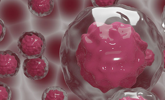
Stem cells are present in all body tissue. They have the capacity to stimulate and proliferate cell growth and can also self-renew. Vesicular secretions from stem cells, called exosomes, create the same effects as the stem cells themselves.
Stem cell transplantation performed on patients rendered sterile after gonadotoxic treatment such as radiation or chemotherapy have in reports, been noticed to sometimes experience return of their fertility. In animal experimentation, stem cells and their exosomes have been shown to mediate return of fertility after egg production has been destroyed. There have been occasional reports of patients with “poor response in IVF” treatments could benefit from this therapy.
At Friberg Fertility, we have been able to improve egg and embryo quality and quantity, and also increase anti-müllerian hormone (AMH) levels in some of our IVF patients who had poor results in earlier IVFs. Exosomes (from mesenchymal stem cells) and platelet-rich plasma (PRP) are injected into the ovaries transvaginally followed by a repeat IVF procedure. After injection, growth factors from exosomes and PRP act on the early stages of egg development and create eggs with a better potential to induce a pregnancy with IVF or intrauterine insemination (IUI). We have applied this approach in our IVF program with success.
Testicular Rejuvenation — Creating Sperm with Stem Cells
In rodents, mesenchymal stem cells (MSC) and stem cell products called exosomes can protect spermatogenetic tissue from destruction after exposure to gonadotoxic treatment. When placed in the right environment – the testicular “niche” — the MSC also develop into mature sperm that have full fertilizing capacity and create embryos. These embryos can then develop to normal fertile offspring.
Direct development of sperm from MSC in humans has not yet been demonstrated, but sperm production has been stimulated in a few azoospermic patients treated with stem cell products. In our approach, we are using a combination of MSC, exosomes and growth-factor rich platelet-rich plasma (PRP) to activate sperm production in men with non-obstructive azoospermia or severe oligozoospermia (cryptozoospermia). The goal is to obtain some sperm in the ejaculate so that the couple can be eligible for an IVF with intracytoplasmic sperm injection (ICSI).
Creating Pregnancies After Menopause
The current treatment of infertility is dependent on the presence of egg cells in the ovaries. At menopause and premature menopause (when menopause occurs before age 40), as well as after radiation and chemotherapy for malignancies, oocytes get destroyed. When only about 1,000 eggs remain in the ovaries, the menstrual cycle stops, but some unstimulated eggs usually remain in the ovary. Why these remaining eggs do not respond is still unclear, but it has something to do with the intraovarian environment or the “ovarian niche.”
Over the past 10 years, the control mechanisms for oocyte development have been revealed. Follicular/oocyte activation follows a pathway called PI-3-AKT and mTOR that promotes cell survival, growth and proliferation. “Hippo” signaling is a system that controls the size of the internal organ. Fragmentation of ovarian tissue inhibits the Hippo signaling. After removal of an ovary or a piece of the ovary and cutting it in small pieces to inhibit the Hippo signaling and stimulate the ovarian pieces with activators and inhibitors of the PI-3-AKT and mTOR pathway, oocytes can be generated from immature oocyte precursors. The treated pieces are then returned to a peritoneal pouch near the fallopian tubes. A variation of the technique with only fragmentation of ovarian tissue (suppression of the Hippo signaling) has also been introduced and resulted in pregnancies. The technique is relatively new, and use of it has resulted in about 20 children being born worldwide.
After institutional review board (IRB) approval, we are planning to apply this “in vitro activation” to stimulate egg production in patients with premature ovarian failure or poor ovarian response/diminished ovarian reserve. The in vitro activation can produce mature eggs for IVF so that these patients may be able to try to have their own biological children which previously was not possible.